Target Audience & Market Research

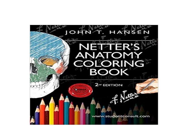
Netters anatomy coloring book – Netter’s Anatomy Coloring Book, leveraging the iconic Netter illustrations, has the potential to be a huge hit, but nailing the target audience and market research is key. We’re not just talking about another anatomy coloring book; we’re talking about a premium product capitalizing on a well-established brand and its reputation for accuracy and detail.This project requires a laser focus on understanding who will buy this book and why.
A comprehensive marketing strategy hinges on this understanding, guiding everything from pricing and distribution to promotional campaigns.
Ideal Age Range and Skill Level
The ideal age range for Netter’s Anatomy Coloring Book spans a broad spectrum, from serious pre-med students to seasoned medical professionals seeking a unique way to reinforce their knowledge. We’re looking at a primary target audience of college-aged students (18-25) enrolled in biology, pre-med, and related courses. However, a significant secondary audience exists among medical professionals (25-55) and even older individuals interested in a detailed and visually engaging anatomy guide.
The skill level should be considered flexible. Beginner-level users might focus on basic coloring and labeling, while advanced users can engage with more intricate details and complex anatomical structures. The book should cater to both groups with varying levels of complexity within its pages.
Potential Market Size and Demographics
The potential market size is substantial. The combined number of pre-med and medical students, healthcare professionals, and individuals interested in human anatomy is considerable. Demographically, the target audience is diverse, encompassing students from various socioeconomic backgrounds and geographic locations. We can anticipate a strong interest in the book from both male and female buyers, reflecting the balanced representation of genders within the medical field and related academic disciplines.
We can use existing sales data for other Netter products, along with market research on anatomy textbooks and educational materials, to project a reasonable estimate for potential sales. For example, if Netter’s Anatomy Atlas sells X number of copies annually, we can reasonably predict a substantial portion of that market would also be interested in a coloring book format.
Netter’s Anatomy Coloring Book offers a detailed approach to learning human anatomy, engaging visual learners through its intricate illustrations. The vibrant, detailed style reminds me of the artistic energy found on the coloring book vinyl Chance the Rapper released a few years back – a completely different subject, of course, but equally captivating in its visual presentation.
Returning to Netter’s, the book’s methodical approach helps solidify understanding of complex structures, making it a valuable tool for students and professionals alike.
This isn’t a shot in the dark; it’s leveraging an existing, proven market.
Comparison to Existing Anatomy Coloring Books
Existing anatomy coloring books often fall into two categories: simplified versions for younger learners and more detailed but less visually appealing books for older students. Netter’s Anatomy Coloring Book differentiates itself by offering the unparalleled detail and accuracy of Netter’s illustrations in a user-friendly coloring book format. This unique combination of high-quality visuals and engaging activity provides a significant competitive advantage.
Think of it as the “high-end” version – like comparing a basic sketchpad to a Moleskine notebook. We’re offering a premium product with a premium price point to match the quality and the brand recognition.
Marketing Strategy
The marketing strategy should focus on highlighting the unique selling proposition: the combination of Netter’s iconic illustrations and the engaging format of a coloring book. This will appeal to both educational institutions and individual consumers. We can leverage social media marketing (Instagram, TikTok) with visually appealing content showcasing the book’s illustrations. Targeted online advertising on platforms frequented by medical students and professionals is also crucial.
Collaborations with relevant influencers (doctors, medical students, science communicators) could boost brand awareness and generate excitement. Additionally, partnerships with bookstores and online retailers are vital for distribution. We should also consider offering bundle deals with other Netter products to encourage purchases. Think of a back-to-school campaign targeted at university bookstores, or collaborations with medical schools for bulk orders.
This isn’t just selling a book; it’s selling access to a renowned brand and a unique learning tool.
Illustrations: Netters Anatomy Coloring Book

Get ready to unleash your inner artist and dive deep into the fascinating world of the human nervous system! We’re talking vibrant colors, detailed anatomical structures, and a whole lotta brainpower. This section will guide you through creating stunning illustrations of the brain, spinal cord, and cranial nerves – think of it as a coloring book upgrade, supercharged with knowledge.
Human Brain Illustration
This illustration showcases a human brain, sliced sagittally (down the middle) to reveal its internal structures. The coloring style is a blend of realistic anatomical accuracy and artistic flair. Think muted earth tones for the deeper brain structures, transitioning to brighter, more saturated colors for the cerebral cortex. For example, the cerebrum is rendered in shades of warm browns and tans, highlighting the gyri and sulci (the folds and grooves) that give it its characteristic wrinkled appearance.
The cerebellum, responsible for coordination and balance, is depicted in cool blues and greens, contrasting beautifully with the cerebrum. Key regions are clearly labeled, including the frontal lobe (executive functions, decision-making), the parietal lobe (sensory processing), the temporal lobe (auditory processing, memory), and the occipital lobe (visual processing). The thalamus, acting as a relay station for sensory information, is shown in a vibrant yellow, while the hypothalamus, regulating vital functions like temperature and hunger, is a deep orange.
The brainstem, connecting the brain to the spinal cord, is depicted in a gradient of purples and reds, emphasizing its crucial role in controlling basic life functions. The corpus callosum, the bridge connecting the two cerebral hemispheres, is a striking white, clearly highlighting its role in interhemispheric communication.
Spinal Cord and Peripheral Nervous System Illustration
This illustration depicts the spinal cord, a long, cylindrical structure extending from the brainstem, encased within the protective vertebral column. The spinal cord is illustrated in a pale, creamy yellow, representing its myelinated nerve fibers. The illustration clearly shows the dorsal (posterior) and ventral (anterior) roots, which merge to form the spinal nerves. These nerves, representing the peripheral nervous system, radiate outwards from the spinal cord, branching into various plexuses and ultimately reaching the muscles and organs of the body.
The peripheral nerves are shown in varying shades of blue and green, depending on their function and location. For example, sensory nerves (carrying information to the CNS) might be a lighter blue, while motor nerves (carrying commands from the CNS) might be a darker green. The illustration also shows the meninges (protective layers surrounding the spinal cord) in subtle shades of grey and pink, adding depth and anatomical accuracy.
The overall style is clean and uncluttered, ensuring clarity and ease of understanding. Think of it like a meticulously drawn subway map, but for your nervous system.
Cranial Nerves
This page utilizes a three-column responsive HTML table to present a clear and organized view of the twelve cranial nerves.
| Cranial Nerve | Origin | Target Area & Function |
|---|---|---|
| Olfactory (I) | Olfactory bulb | Nasal mucosa; sense of smell |
| Optic (II) | Retina | Visual cortex; vision |
| Oculomotor (III) | Midbrain | Eye muscles; eye movement |
| Trochlear (IV) | Midbrain | Superior oblique muscle; eye movement |
| Trigeminal (V) | Pons | Face, mouth, and jaw; sensation and chewing |
| Abducens (VI) | Pons | Lateral rectus muscle; eye movement |
| Facial (VII) | Pons | Facial muscles; facial expression, taste |
| Vestibulocochlear (VIII) | Pons and medulla | Inner ear; hearing and balance |
| Glossopharyngeal (IX) | Medulla | Tongue, pharynx; swallowing, taste, salivation |
| Vagus (X) | Medulla | Thorax and abdomen; parasympathetic control |
| Accessory (XI) | Medulla and spinal cord | Neck and shoulder muscles; head and shoulder movement |
| Hypoglossal (XII) | Medulla | Tongue muscles; tongue movement |
Illustrations: Netters Anatomy Coloring Book

Get ready to unleash your inner artist and bone up on your anatomy knowledge! This section dives deep into the illustrations of the musculoskeletal system, bringing the human body to life, one vibrant color at a time. We’re talking seriously detailed visuals, the kind that’ll make your textbook illustrations look like stick figures. Think of it as a backstage pass to the body’s most impressive stage show – the intricate dance of bones and muscles.
Human Skeleton Illustration: Bone Markings and Articulations, Netters anatomy coloring book
This detailed illustration showcases the complete human skeleton, highlighting key bone markings and articulations. Imagine a breathtaking panoramic view of the skeletal system. Each bone is meticulously rendered, revealing its unique shape and size. Notice the subtle curves of the spine, the delicate structure of the hand bones, and the robust architecture of the femur. Key bone markings, like the greater trochanter of the femur (where powerful hip muscles attach), the glenoid fossa of the scapula (the socket for the shoulder joint), and the various processes and foramina (openings for nerves and blood vessels), are clearly labeled and highlighted with color-coding to emphasize their functional significance.
The articulations, or joints, are shown with special attention to their structure, illustrating how bones fit together to allow for a wide range of movements. The temporomandibular joint (TMJ), the knee joint with its complex ligaments, and the intricate ball-and-socket hip joint are all depicted in stunning detail, showcasing the amazing engineering of the human body. Think of it like a detailed architectural blueprint of your body’s framework.
Major Muscle Groups Illustration: Origins, Insertions, and Actions
This illustration depicts the major muscle groups of the body, emphasizing their origins, insertions, and actions. Picture a powerful, sculpted figure, with each muscle group meticulously defined and colored. The illustration clearly shows the origin (the point of attachment of a muscle to a more stationary bone) and insertion (the point of attachment to a more movable bone) of key muscles.
For example, the biceps brachii muscle’s origin is highlighted on the scapula, and its insertion on the radius, clearly demonstrating how contraction of this muscle causes flexion of the elbow. The illustration also details the actions of each muscle group – flexion, extension, abduction, adduction, rotation, etc. – providing a clear visual understanding of how these muscles work together to produce movement.
This illustration is your ultimate cheat sheet to understanding how your muscles power your every move, from the subtle twitch of an eyelid to the powerful lunge of a basketball player.
Upper and Lower Limb Muscles and Bones
This section uses a responsive four-column table to provide a detailed look at the bones and muscles of the upper and lower limbs. Think of it as a super-organized, easy-to-navigate anatomy cheat sheet.
| Upper Limb – Bone | Upper Limb – Muscle | Lower Limb – Bone | Lower Limb – Muscle |
|---|---|---|---|
| Humerus (detailed view showing deltoid tuberosity, etc.) | Biceps Brachii (origin, insertion, action clearly labeled) | Femur (detailed view showing greater trochanter, etc.) | Quadriceps Femoris (rectus femoris, vastus lateralis, vastus medialis, vastus intermedius – origins, insertions, actions) |
| Radius & Ulna (articulation clearly shown) | Triceps Brachii (origin, insertion, action clearly labeled) | Tibia & Fibula (articulation clearly shown) | Hamstrings (biceps femoris, semitendinosus, semimembranosus – origins, insertions, actions) |
| Carpals, Metacarpals, Phalanges (detailed view of articulations) | Flexor Carpi Radialis (origin, insertion, action clearly labeled) | Tarsals, Metatarsals, Phalanges (detailed view of articulations) | Gastrocnemius & Soleus (origin, insertion, action clearly labeled) |
Clarifying Questions
What age group is this coloring book suitable for?
It’s suitable for a wide range, from high school students to medical professionals, adapting to different skill levels and knowledge bases.
Are the illustrations medically accurate?
Yes, the illustrations are based on Netter’s renowned anatomical accuracy, simplified for clarity and learning.
Can this book be used as a supplemental learning tool for medical students?
Absolutely! It complements traditional learning methods by enhancing memorization and visual understanding.
What kind of paper is used in the book?
High-quality paper is used to prevent bleed-through and allow for layering of colors.


